Process for enriching erythroblasts
- Erythroblasts are known as fetal cells which enter the maternal circulation through the placenta. The existent ratio of erythroblasts is significantly lower than maternal leukocytes, and is considered as less than 1/10
 maternal nucleated cells.
 - Majority of maternal erythrocytes is first purged by 1.095 g/mL single density gradient centrifugation from maternal blood (processing time: 30 min).
- Following the density gradient centrifugation, CD45 depletion using panning dish immobilizing monoclonal antibodies is performed to reduce leukocytes (processing time: 30 min).
- Unattached cells after the panning are recovered, resuspended in PBS supplementing with SBA lectin, and then incubated in the chamber slides modified with G-coat involving galactose (processing time : 45 min).
- The processes described above are throughout performed at room temperature.
- After removing the chamber, the slide glass is subject to centrifugation to fix and dry the attached cells.
- Following additional air-drying, the cells on the slide glasses are stained by May-Grunwald Giemsa (MGG) solution to identify morphological erythroblasts.
- Subsequently, genomes of fetal erythroblasts are examined.
Detection of isolated erythroblasts
- Fetal and maternal erythroblasts coexist in maternal circulation. Since fetal and maternal erythroblasts are the same nature, the lectin method preferentially attaches both erythroblasts without discrimination.
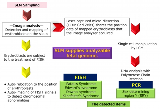
- Cells which were dried and fixed following the isolation by lectin method are stained with MGG, and then microscopically identified. Representative erythroblasts having wide cytoplasm and dark-colored nucleus are shown below. In our study, erythroblasts were efficiently detected by image analyzer, Metafer4/Axio (Carl Zeiss).
- Operator selects the representative erythroblasts from the presented cells, and then the selected position data is stored in the image analyzer.
- We could detect 18 to 240 erythroblasts from 10 mL maternal blood which was collected from pregnant women within 18 weeks.
Cytogenetic assay of fetal erythroblasts by using commercially available image analyzer that we actually have done is shown below.

SBA-lectin method (SLM) supplies analyzable fetal genome.

 Contact US

