Related informations of Lectin method
2021-10-12
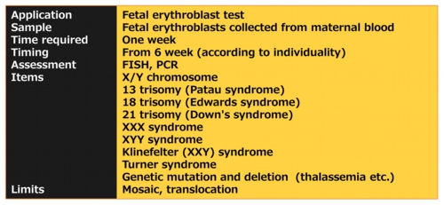
Fetal erythroblast test

Problems of prenatal tests
1. Profile of definitive diagnosis including invasive sampling of amniocytes and chorionic villus (CVS)
- Amniotic fluid containing amniocytes of fetal tissues is collected from the amnion or amniotic sac surrounding a developing fetus.
- In order to collect a villi including fetal tissue cells, a small needle/catheter is placed either through the abdomen or through the vagina near the uterus.
- Invasive collections of fetal cells are occasionally accompanied by miscarriage or deformation of fetus.
- Analysis of fetal nucleated cells leads to the definitive diagnosis from which the fetal chromosome and DNA is directly examined.
2. Profile of non-invasive probability test
- The maternal serum test is a blood test without miscarriage risk for pregnant women in the first or second trimester of pregnancy. The serum test helps imply the risk of certain abnormalities such as Down syndrome, Edward syndrome and spina bifida. The test does not indicate whether congenital anomalies are definitely present. Test results contain false negative and positive.
- If the test suggests an doubtable risk, the pregnant woman can then decide whether to undertake further definite diagnosis such as an amniocentesis or chorionic villus sampling (CVS).
- Several serum components in maternal blood such as β-hCG, inhibin-A, estriol, h-hCG are measured, and then the pattern of those concentrations is obtained.
- The obtained patterns is compared with the statistical pattern of no-risk pregnancy. A gap among the patterns is indicative of the risks of chromosomal abnormalities.
Erythroblast
- Erythroblast is immature red blood cells and also called nucleated red blood cell (NRBC). Fetal whole genome is stored in its nucleus.
- Placenta consists of maternal decidua basialis and fetal chorion frondosum. Fetal chorion frondosum merges with maternal decidua basialis which fills with maternal venous blood, to take up oxygen and nutrition. Active metabolism occurs on the fetal area, thereby fetal cells such as trophoblastic cells continuously repeats degradation and reproduction during the period of pregnancy. This turnover produces fetal DNA fragments based on the degradation. Consequently these DNA fragments enter maternal circulation, and then become the source of cell-free DNA testing.
- It is presumed that undegraded cells, such as lymphocyte or the erythroblast as well as trophoblastic cell, enter maternal venous blood through such process, but its frequency is considerably low. It is known that hypertension pregnancy arising from various causes, so-called preeclampsia, increases the number of erythroblasts in maternal venous blood whereas the entering mechanism of cells which are considerably large particle than DNA fragments is not completely clarified.
- Reason why erythroblasts become the target of prenatal test concerns the life span of blood cells. For example, lymphocyte's life span is several years, therefore, fetal lymphocytes in maternal blood cannot deny the possibility of former pregnancy origin. In other words, it is the possibility of older brother/sister origin. In contrast, erythroblast's life span is less than a year, thus there is no anxiety for the prenatal test.
- From the profile of blood cells described below, the isolation of erythroblasts from leukocytes turns out to be difficult in terms of the insignificant difference of size between leukocytes and erythroblasts. In addition, low existence ratio of erythroblasts is the substantial difficulty for precise isolation.


Comparison of prenatal test
- Accuracy of the cell free DNA test is significantly higher than that of the maternal serum marker test.
- But the cost of the cell free DNA is more than 10 times of that of the serologic marker.
- Erythroblast test is a unique examination without the miscarriage risk despite bringing a definite result.
- Single cell manipulation of erythroblasts having fetal whole genome enables to simplify DNA analysis.

Fluorescence in situ Hybridization (FISH)
- FISH involves the hybridization of fluorescent oligonucleotide probes to their complementary chromosome of cells on the specimens. Recently Silver-ISH to enzymatically develop black coloration is also used for detecting the breast cancer cells. The most important feature of ISH is to enable microscopically visual detection of a specific region of the chromosomes. The visual detection becomes the powerful methodology for detecting the chromosomal anomalies such as not only aneuploidy but also Translocation and Mosaic.
- In addition, it is revealed that genetic point mutation or translocation of locus modulating cell proliferation induces constant activation namely carcinogenesis. In recent years, it was reported that minute translocation of the chromosome 2 occurs in non‐small cell lung cancer. In the normal case, red- and yellow-spot signals are individually observed at a distant place, when two loci on chromosome which can be fused through translocation hybridize to their FISH probes with red- or green-fluorescent labeling. In the abnormal case with carcinogenesis, yellow-spot signals are observed through fusing two loci including red and green. This sort of analytical method for the translocation is expected as a new detection method of carcinogenesis.
- FISH is the analytical method that depends on an expert technique than DNA analysis using PCR (Polymerase Chain Reaction) which mechanical automation goes ahead through. The basic process of FISH includes (1) dehydration and fixation, (2) aging with surfactants, (3) bare nucleus with the enzyme, (4) fixation, (5) denaturation of subject DNA and its probe, (6) hybridization, (7) removal of non-specific fluorescence, and (8) counter stain with DAPI. Subsequently FISH asks for optimization of each process according to cellular type and state of the specimens.
"NeTech has optimized FISH process for erythroblasts arising from maternal blood."
Principle of PCR
- PCR (Polymerase chain reaction) is a technique that extremely contributed to advance of modern molecular biology. Its technological innovation and the price reduction of its consumables is advancing with an astonishing speed. PCR enables to expeditiously amplify a specific locus from a small amount of DNA specimen, and then to precisely detect the amplified loci. The PCR is nowadays applied to wide fields, such as clinically genetic testing, food hygiene test, crime investigation, as well as fundamental researches, by reason of its serviceability.
- Genetic analysis using PCR involves various kinds of processes depending on the detection method of the copies. Real time PCR (RT-PCR) is a general-purpose technology and is performed according to the process describing below (please refer to a book or an interpretation on network, etc. for details).
- 1. DNA extraction
- DNA is enzymatically extracted from histopathology or isolated cells according to the manual of commercially available products. The products without the extraction process are also commercialized.
- 2. Amplification of DNA
- Fundamentally, DNA amplification is achieved by repetition of a) denaturation, b) annealing, and c) polymerization.
- Denaturation: Specimen is heated at 94 ℃ to denature DNA. The denaturation turns double strand DNA into the single.
- Annealing: In order to activate DNA polymerase, annealing for combining a primer (DNA template) is performed beforehand. Generally the primer complementarily combines with the end of the specific locus at 55~60 ℃. Depending on the primer used, the temperature besides this range may be adopted.
- Polymerization: The polymerase reacts with the end of the primer, extends DNA sequence, and then produces the copies of the specific locus. In the case to use heat-resistant Taq Polymerase, approximately 72 ℃ is adopted.
- By repeating the above process with dedicated equipment, thermal cycler, the specific locus is amplified exponentially. Since the specific locus increases twofold by the one thermal cycling, the twenty two cycles numerically produces one million copies. In actuality, about 30 cycles are carried out. Consequently, it will be amplified within the range from 1 to 10 million times.
3.Detection of reproduced DNA
iiiii) Fluorescent DNA intercalator
- The fluorescent intercalators which are applied during PCR combine to the reproduced double strand DNA, and then emit the fluorescence by irradiating excitation light. The fluorescence intensity continuously increases depending on the reproduction of DNA..
iiiii) TaqMan fluorogenic probe
- TaqMan probe, that a fluorescent substance (FAM and the like) and a quencher (TAMRA and the like) is introduced at the 5' end and the 3' end on an oligonucleotide, are applied to the process of PCR . Though TaqMan probe hybridizes to the primer through annealing, the quencher inhibits the probe from emitting.
- During elongation step following the annealing, 5′→3′ exonuclease activity of Taq DNA protease decomposes TaqMan probe which hybridizes to the primer, and then the fluorescent substances are dissociated from the probe fragment including the quencher. Finally the dissociation from the quencher enables to emit fluorescence.
iiiii) Cycling probe technology (CPT)
- CPT is a precise detection method by means of chimeric DNA-RNA-DNA probe combining the fluorescent reporter together with its quencher, which is cleaved by RNase H to selectively cut within the RNA portion. The enzyme does not cut single-stranded DNA or RNA.
- The formation of double-stranded DNA is carried out at a temperature that allows the chimeric probe to anneal to the single-stranded target DNA. After cutting the chimeric probe by RNase H, the shorter fragments dissociate from the target DNA. The formation of double-stranded DNA and the enzymatic fragmentation is continuously repeated on the target DNA after the dissociation.
- This cycling reaction produces abundant fluorescent reporters from a slight target DNA and enables low background and precise detection. In addition, CPT is fast, linear, isothermal and simple than other DNA detection methods.
General-purpose PCR
- RT-PCR is also called quantitative PCR (qPCR) and is one of the most practical technologies for genetic assessment. Therefore it is used in wide fields such as quantification of gene expression, genotyping, single nucleotide polymorphism (SNP), oncogene detection, confirmation of drug efficacy, the measurement of RNA interference, and the like.
- RT-PCR is a method of determining in real time an amount of DNA as augmentation of fluorescence intensity corresponding to DNA amplification. Consequently RT-PCR serves as the reverse transcription to quantify messenger RNA (mRNA) and microRNA (miRNA). The simpleness of RT-PCR drastically expanded the application of genetic analysis involving PCR. On the other hand, RT-PCR demands the calibration curve based on an fluorescent standard substance and thereby produces the measurement errors.
- In the first place, traditional PCR accumulates the reproduced substances after finishing PCR, and semi-quantitatively measures them. Therefore, the estimation of the quantitative changes concerning the amplified nucleic acids will be difficult.
- Digital PCR (d-PCR) following the conventional PCR technologies will be clearly differentiated from them. d-PCR is q-PCR intrinsically, but shows overwhelming ability for the absoluteness of quantification and the detection of a rare allele. Digital-PCR enables to directly count the number of DNA fragments which was copied. Consequently d-PCR will achieve objective quantification which does not rely upon the calibration curve based on a standard material and the comparison with amplification of an internal standard gene.
Digital PCR
- Digital-PCR enables the absolute quantification of copied DNA as well as the detection of infrequently occurring mutation in DNA sequence and of copy number variation (CNV). The deep sequencing through next-generation sequencer is also applicable to analysis for one molecule DNA. Comparison between d-PCR and next-generation sequencing is described below.
- On the detection of infrequently occurring mutation, an error due to the amplification and the failure of identifying nucleic acid sequence becomes a substantial problem at next-generation sequencer. In this point, the sequencer is inferior to digital PCR apparatus.
- The sequencer is hard to precisely detect CNV.
- The sequencer is hard to absolutely quantify target DNA.
- The disadvantage of Digital PCR instrument in the detection of mutant DNA is to be limited to the analysis of specific DNA mutations. Thus d-PCR is not suitable for the screening of genetic mutation or deletion.
Principle of d-PCR
- One molecule of the copy is reproduced by thermal cycler in the microchambers or nano/picoliter of droplets, and then TaqMan probe of the copy radiates fluorescence. Digitally checking the fluorescence of each chamber or droplet unit results in absolute quantification. In addition, the reaction in limited and cramped space suppresses the occurrence of errors, and then drastically improves the accuracy of detection. Two-color probe which enables multi-item examination has also been commercialized from some manufacturers.
- It is expected that d-PCR becomes a powerful technology to cytogenetically test erythroblasts less than 100 cells, as well as FISH.

 Contact US
 Home
Home Related informations